推荐厂家
暂无
暂无


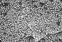
我在JEM2010(高分辨)上照的8000倍的图像,算不算高分辨图像,
[align=center][font='times new roman'][size=16px][b]超高分辨[/b][/size][/font][font='times new roman'][size=16px][b]显微镜及其在生物医学领域的应用[/b][/size][/font][/align][align=center][font='times new roman'][size=14px]刘皎[/size][/font][font='times new roman'][sup][size=14px]1[/size][/sup][/font][font='times new roman'][size=14px],[/size][/font][font='times new roman'][sup][size=14px] [/size][/sup][/font][font='times new roman'][size=14px]吴晶[/size][/font][font='times new roman'][sup][size=14px]1[/size][/sup][/font][/align][align=center]1. [font='times new roman']北京大学医药卫生分析中心,北京,[/font][font='times new roman']100191[/font][/align][font='times new roman'][b]摘要[/b][/font][font='times new roman'][b] [/b][/font][font='times new roman']超高分辨显微镜([/font][font='times new roman']Super-Resolution Microscopy[/font][font='times new roman'])作为一类强大的科学工具,可以突破传统光学显微镜的分辨极限,实现对微小结构的高分辨率成像,已经在生物医学领域引起了广泛的关注和应用。本文将探讨超高分辨显微镜的不同类型和原理,介绍[/font][font='times new roman']其[/font][font='times new roman']在生物医学领域的应用[/font][font='times new roman']及展望其未来发展[/font][font='times new roman']。[/font][font='times new roman'][b]Abstract[/b][/font][font='times new roman']Super Resolution Microscopy[/font][font='times new roman'], as a powerful scientific tool, can break through the resolution limit of traditional optical microscopes and achieve high-resolution imaging of small structures. It has attracted widespread attention and application in the biomedical field. This article will explore the different types and principles of Super Resolution Microscopy, introduce their applications in the biomedical field, and look forward to their future development[/font][font='times new roman'].[/font][font='times new roman'][b]关键词[/b][/font][font='times new roman']超高分辨[/font][font='times new roman']显微镜,[/font][font='times new roman']成像技术[/font][font='times new roman'],应用[/font][font='times new roman'][b]1 [/b][/font][font='times new roman'][b]引言[/b][/font][font='times new roman']显微镜的产生和发展对于生命科学研究的进步有至关重要的作用[/font][font='times new roman'],它将微观世界呈现在大家面前,包括微生物的存在、组织细胞结构及生理病理活动等。显微镜技术的不断革新将成像分辨率不断提高,但相当长一段时间内光学成像无法突破一个极限值,即[/font][font='times new roman']xy[/font][font='times new roman']轴横向分辨率约[/font][font='times new roman']200nm[/font][font='times new roman'],[/font][font='times new roman']z[/font][font='times new roman']轴纵向分辨率约[/font][font='times new roman']500nm[/font][font='times new roman'],因此小于这个尺寸的生命活动和结构[/font][font='times new roman'],如病毒、亚细胞结构等,[/font][font='times new roman']是无法清楚地观察到的[/font][font='times new roman']。[/font][font='times new roman']聚焦点的光强会根据点扩散函数([/font][font='times new roman']point spread functio[/font][font='times new roman']n[/font][font='times new roman'],[/font][font='times new roman']PSF[/font][font='times new roman'])而展开[/font][font='times new roman'],[/font][font='times new roman']对于圆形孔径,[/font][font='times new roman']PSF[/font][font='times new roman']呈现为艾里斑([/font][font='times new roman']Airy disk[/font][font='times new roman'])的模式[/font][font='times new roman']。[/font][font='times new roman']激光扫描共聚焦显微镜([/font][font='times new roman']Confocal Laser Scanning Microscopy, CLSM[/font][font='times new roman'])的分辨率取决于[/font][font='times new roman']PSF[/font][font='times new roman']的大小,如果焦点很小,则每个像素[/font][font='times new roman']点[/font][font='times new roman']获取到的信息也很小,从而得到清晰锐利的图像;反之,则结果图像变得模糊。因此,[/font][font='times new roman']CLSM[/font][font='times new roman']成像的[/font][font='times new roman']主要挑战在于实现越来越小的[/font][font='times new roman']PSF[/font][font='times new roman']以获得更好的分辨率。德国物理学家恩斯特[/font][font='times new roman'][/font][font='times new roman']阿贝([/font][font='times new roman']Ernst Abbe[/font][font='times new roman'],[/font][font='times new roman']1840-1905[/font][font='times new roman']年)在[/font][font='times new roman']19[/font][font='times new roman']世纪[/font][font='times new roman']70[/font][font='times new roman']年代首次[/font][font='times new roman']提出阿贝衍射极限,即[/font][font='times new roman']由于衍射效应,[/font][font='times new roman']PSF[/font][font='times new roman']大[/font][font='times new roman']小与[/font][font='times new roman']λ/NA[/font][font='times new roman']成正比([/font][font='times new roman']d=0.61λ/NA[/font][font='times new roman']),其中[/font][font='times new roman']λ[/font][font='times new roman']是光的波长,[/font][font='times new roman']NA[/font][font='times new roman']是物镜最重要的参数[/font][font='times new roman']——[/font][font='times new roman']数值孔径[/font][font='times new roman']。由于可见光波长范围在[/font][font='times new roman']400-760nm[/font][font='times new roman']之间,[/font][font='times new roman']NA[/font][font='times new roman']值最大在[/font][font='times new roman']1.7[/font][font='times new roman']左右,所以分辨率极限在[/font][font='times new roman']200nm[/font][font='times new roman']左右。随着物理学和测量技术的进步,突破衍射极限的显微镜不断涌现,目前公认的超高分辨显微镜主要有三类,包括[/font][font='times new roman']结构照明显微镜([/font][font='times new roman']Structured Illumination Microscopy[/font][font='times new roman'],[/font][font='times new roman']SIM[/font][font='times new roman'])[/font][font='times new roman'],受激发射减耗显微镜([/font][font='times new roman']Stimulated Emission Depletion Microscopy[/font][font='times new roman'],[/font][font='times new roman']STED[/font][font='times new roman']),和[/font][font='times new roman']单分子定位显微镜。单分子定位显微镜包括光敏定位显微镜([/font][font='times new roman']Photoactivation Localization Microscopy[/font][font='times new roman'],[/font][font='times new roman']PALM[/font][font='times new roman'])和随机光学重建显微镜([/font][font='times new roman']Stochastic Optical Reconstruction Microscopy[/font][font='times new roman'],[/font][font='times new roman']STORM[/font][font='times new roman'])[/font][font='times new roman']。[/font][font='times new roman']2014[/font][font='times new roman']年三位科学家[/font][font='times new roman']史蒂芬[/font][font='times new roman'][/font][font='times new roman']霍尔([/font][font='times new roman']Stefan W. Hell[/font][font='times new roman'])[/font][font='times new roman']、埃里克[/font][font='times new roman'][/font][font='times new roman']贝兹([/font][font='times new roman']Eric Betzig[/font][font='times new roman'])和威廉[/font][font='times new roman'][/font][font='times new roman']莫纳([/font][font='times new roman']William E. Moerner[/font][font='times new roman'])因他们在超[/font][font='times new roman']高[/font][font='times new roman']分辨显微镜技术领域的贡献而获得了诺贝尔化学奖。[/font][font='times new roman'][b]2 [/b][/font][font='times new roman'][b]不同类型的超高分辨显微镜[/b][/font][font='times new roman'][b]2.1[/b][/font][font='times new roman'][b] [/b][/font][font='times new roman'][b]结构照明显微镜([/b][/font][font='times new roman'][b]Structured Illumination Microscopy[/b][/font][font='times new roman'][b],[/b][/font][font='times new roman'][b]SIM[/b][/font][font='times new roman'][b])[/b][/font][font='times new roman']SIM[/font][font='times new roman']本质是利用两束激发光在样品上进行干涉,产生明暗交替的莫尔条纹,高空间频率的莫尔条纹会放大激发条纹与样品空间频率不一致的结构,从而将样品中的高频信息整合入收集到的图像中。[/font][font='times new roman']通过投射特殊的光照明模式如格点或条纹光栅,以一定的模式照射样品,引入空间频率信息,采集多个图像并经过复杂的数据处理之后,重建高分辨率图像。由于每个图像都采用不同的结构照明模式,包含了不同的信息,合并后的图像能够展示出比传统显微镜更多的细节[/font][font='times new roman']。[/font][font='times new roman']相比于其他超高分辨成像技术,[/font][font='times new roman']SIM[/font][font='times new roman']最大的优势就是宽场[/font][font='times new roman']成像,速度快,基本可以达到实时观察。[/font][font='times new roman']SIM[/font][font='times new roman']技术的前身可以追溯到[/font][font='times new roman']20[/font][font='times new roman']世纪[/font][font='times new roman']70[/font][font='times new roman']年代初。当时,光学学家特奥多尔[/font][font='times new roman'][/font][font='times new roman']赫普恩([/font][font='times new roman']Theodor [/font][font='times new roman']H?upl[/font][font='times new roman'])首次提出了使用周期性光栅照明来提高显微镜分辨率的想法。这奠定了[/font][font='times new roman']SIM[/font][font='times new roman']技术的基础,尽管当时还没有实际的[/font][font='times new roman']SIM[/font][font='times new roman']显微镜。[/font][font='times new roman']21[/font][font='times new roman']世纪初期,史蒂芬[/font][font='times new roman'][/font][font='times new roman']霍尔([/font][font='times new roman']Stefan W. Hell[/font][font='times new roman'])和埃里克[/font][font='times new roman'][/font][font='times new roman']贝兹([/font][font='times new roman']Eric Betzig[/font][font='times new roman'])等科学家分别独立开发了[/font][font='times new roman']SIM[/font][font='times new roman']的现代版本。[/font][font='times new roman']SIM[/font][font='times new roman']技术开始广泛传播,吸引了生物学家和显微镜专家的关注。它被认为是一种相对低成本的[/font][font='times new roman']超高分辨[/font][font='times new roman']率成像方法,因为它不需要昂贵的激光设备或复杂的样品准备。[/font][font='times new roman'][b]2.2 [/b][/font][font='times new roman'][b]受激发射减耗[/b][/font][font='times new roman'][b]显微镜([/b][/font][font='times new roman'][b]Stimulated Emission Depletion Microscopy[/b][/font][font='times new roman'][b],[/b][/font][font='times new roman'][b]STED[/b][/font][font='times new roman'][b])[/b][/font][font='times new roman']STED[/font][font='times new roman']技术的概念最早由斯德哥尔摩大学的斯蒂芬[/font][font='times new roman'][/font][font='times new roman']霍尔([/font][font='times new roman']Stefan W. Hell[/font][font='times new roman'])提出。他的想法是通过将激发光束与一个特殊的抑制光束结合,从而实现对荧光标记物的光抑制,[/font][font='times new roman']通过受激辐射淬灭光斑外围的荧光分子,[/font][font='times new roman']使其在空间上变得更加紧凑,[/font][font='times new roman']减少[/font][font='times new roman']PSF[/font][font='times new roman']从而提高分辨率。[/font][font='times new roman']我们也叫“甜甜圈”技术。[/font][font='times new roman']STED[/font][font='times new roman']显微镜背后基本思想就是利用非线性光学设计一个低于阿贝衍射极限的更小[/font][font='times new roman']PSF[/font][font='times new roman']。[/font][font='times new roman']分辨率与[/font][font='times new roman']STED[/font][font='times new roman']光强有关,提高[/font][font='times new roman']STED[/font][font='times new roman']光的强度可以使荧光光斑焦[/font][font='times new roman']点中心直径趋于[/font][font='times new roman']0[/font][font='times new roman'],但是实际应用中,光损伤较大,[/font][font='times new roman']STED[/font][font='times new roman']光强不可能无限增加,顾[/font][font='times new roman']其分辨率[/font][font='times new roman']最高[/font][font='times new roman']可达到[/font][font='times new roman']3[/font][font='times new roman']0[/font][font='times new roman']nm[/font][font='times new roman']左右[/font][font='times new roman']。[/font][font='times new roman']目前的[/font][font='times new roman']STED[/font][font='times new roman']只能应用于较薄的组织器官或细胞,光毒性较强,成像厚度有限不太适合活体或活细胞长时间成像。[/font][font='times new roman']STED[/font][font='times new roman']光路较为复杂,对系统稳定性要求较高。[/font][font='times new roman'][b]2.3 [/b][/font][font='times new roman'][b]单分子定位显微镜[/b][/font][font='times new roman']单分子定位显微镜[/font][font='times new roman']中荧光标记的单个分子被分别激发和检测。单分子的中心可以以极高的精度确定从而实现高分辨率,包括光敏定位显微镜([/font][font='times new roman']Photoactivation Localization Microscopy[/font][font='times new roman'],[/font][font='times new roman']PALM[/font][font='times new roman'])和随机光学重建显微镜([/font][font='times new roman']Stochastic Optical Reconstruction Microscopy[/font][font='times new roman'],[/font][font='times new roman']STORM[/font][font='times new roman'])。[/font][font='times new roman']PALM[/font][font='times new roman']的历史可以追溯到[/font][font='times new roman']2006[/font][font='times new roman']年,由埃里克[/font][font='times new roman'][/font][font='times new roman']贝兹([/font][font='times new roman']Eric Betzig[/font][font='times new roman'])和哈拉尔德[/font][font='times new roman'][/font][font='times new roman']赫斯([/font][font='times new roman']Harald Hess[/font][font='times new roman'])提出了单分子定位这一概念。在[/font][font='times new roman']PALM[/font][font='times new roman']中,样品中的分子被标记上特定的荧光染料。这些染料可以通过光激活从一个基态转变到一个激发态,此过程可通过使用激活光(通常是紫外光)来实现。同期[/font][font='times new roman']STORM[/font][font='times new roman']的成像技术也发展起来,代表科学家是华人庄小威。[/font][font='times new roman']STORM[/font][font='times new roman']的工作原理与[/font][font='times new roman']PALM[/font][font='times new roman']类似,是通过特殊的分子标记和随机活性化,实现单分子定位进而实现超高分辨率成像。具有光激活能力的标记物通常在某种光照条件下会发光,但也会在某一时刻被随机地熄灭。这种随机光熄灭是[/font][font='times new roman']PALM[/font][font='times new roman']技术的关键,因为它允许在不同时间点捕获标记物的位置。通过记录标记物的位置,可以得到它们的坐标。这一过程需要在短时间内多次拍摄样品,以获得足够多的数据点。最后,通过将多个标记物的坐标叠加在一起,可以生成高分辨率的图像。这种以成像时间换取空间分辨率的形式,使得[/font][font='times new roman']PALM[/font][font='times new roman']或[/font][font='times new roman']STORM[/font][font='times new roman']的分辨率通常能够达到数十纳米。[/font][font='times new roman'][b]3 [/b][/font][font='times new roman'][b]应用领域和未来发展[/b][/font][font='times new roman']超高分辨显微镜可以探索微观世界的无限可能性,已经彻底改变了科学研究的方式。在细胞生物学领域,它被用于研究[/font][font='times new roman']亚细胞结构,如微丝、微管、肌动蛋白等,[/font][font='times new roman']细胞器[/font][font='times new roman']如线粒体、溶酶体等,[/font][font='times new roman']分子分布和细胞膜动态、观察蛋白质的相互作用;在神经科学领域,它可用于观察神经元的亚细胞结构和突触的细节,有助于解剖和理解神经系统的结构和功能,以及神经系统相关疾病的机制;在癌症研究领域,被用于研究癌细胞的特征、蛋白质分布以及肿瘤微环境,这对于癌症的早期诊断和治疗规划非常重要;在材料科学领域,它被用于研究纳米材料的结构和性质、帮助科学家精确控制和制备纳米结构;在药物研发领域,它可用于研究药物靶标蛋白的定位和与其他分子的相互作用,这对于药物设计和筛选非常重要[/font][font='times new roman'];在微生物领域,对于研究细菌[/font][font='times new roman']结构变化至关重要,规避了电子显微镜无法进行活体成像等弊端,可以更加推进微生物学发展。[/font][font='times new roman']当然,[/font][font='times new roman']超[/font][font='times new roman']高[/font][font='times new roman']分辨成像技术[/font][font='times new roman']也有一定的挑战。超高分辨成像技术[/font][font='times new roman']通常需要高度复杂的设备和精密的校准,这使得其设备成本相对较高,[/font][font='times new roman']再加上样本制备的困难,[/font][font='times new roman']限制了其广泛应用。[/font][font='times new roman']样品准备在超高分辨成像中具有重要作用,新的标记技术和荧光探针的发展将提高成像的灵敏度和特异性[/font][font='times new roman'],[/font][font='times new roman']开发更友好、无损伤的样品准备方法,以减少对样品的干扰[/font][font='times new roman'],[/font][font='times new roman']甚至[/font][font='times new roman']包括无标记成像技术以减少样品标记的需求。开源软件和自动化工作流程将使超高分辨成像技术更易于使用和共享,促进科学研究的进展。[/font][font='times new roman']超高分辨技术通常对于三维成像和大样本的深度成像有限制,需要克服分辨率和深度之间的权衡。[/font][font='times new roman']同时超高分辨[/font][font='times new roman']成像的时间分辨率还可以继续提升[/font][font='times new roman']。[/font][font='times new roman']虽然[/font][font='times new roman']目前[/font][font='times new roman']SIM[/font][font='times new roman']和[/font][font='times new roman']minflux[/font][font='times new roman']更适合[/font][font='times new roman']观察[/font][font='times new roman']活细胞[/font][font='times new roman']动态过程,但时间分辨率的提高仍然是一个挑战,特别是对于极短时间尺度的现象[/font][font='times new roman'],[/font][font='times new roman']这将使科学家能够更深入地探索微观世界,并获得更多信息。[/font][font='times new roman']随着技术的不断进步,[/font][font='times new roman']超高分辨[/font][font='times new roman']成像有望在[/font][font='times new roman']包括临床医学[/font][font='times new roman']等[/font][font='times new roman']更多领域得到广泛应用[/font][font='times new roman'],未[/font][font='times new roman']来如果能实现超高分辨的动物甚至人的[/font][font='times new roman']活体成像,减少样品固定和处理的需求,允许观察生物过程的实时发生[/font][font='times new roman']将会更有现实意义[/font][font='times new roman']。[/font][font='times new roman']并且在科学研究的需求下,[/font][font='times new roman']多模态[/font][font='times new roman']或多尺度[/font][font='times new roman']成像将[/font][font='times new roman']与[/font][font='times new roman']不同[/font][font='times new roman']的[/font][font='times new roman']超高分辨[/font][font='times new roman']技术相结合,[/font][font='times new roman']例如,结合光学成像和质谱成像[/font][font='times new roman'],[/font][font='times new roman']从分子水平到组织水平[/font][font='times new roman']提供[/font][font='times new roman']生命活动[/font][font='times new roman']更全面的信息。[/font][font='times new roman']也可以[/font][font='times new roman']发展高通量的样品处理和成像技术,以便更快速地获得大规模的数据。[/font][font='times new roman']超高分辨[/font][font='times new roman']成像生成的数据量巨大,处理和分析这些大数据需要强大的计算资源和高效的算法。数据存储和传输也是挑战。[/font][font='times new roman']超高分辨[/font][font='times new roman']成像数据可能受到噪声和伪迹的影响,这需要高级的图像处理技术来减少其影响,以获得准确的图像。数据分析通常需要复杂的算法和数学模型,需要专业知识和技能。对于某些应用,如神经科学中的活体成像,需要实时数据分析,这增加了挑战。深度学习和人工智能技术[/font][font='times new roman']有望[/font][font='times new roman']在数据分析中发挥越来越重要的作用,[/font][font='times new roman']实现[/font][font='times new roman']自动处理和解释图像数据。发展实时数据分析技术,使科学家能够在数据采集过程中获得及时反馈。开发更易用的高级图像处理工具,使非专业用户也能够进行数据分析。结合不同成像技术和数据源的信息,以提供更全面的信息。开发自动化和高通量的数据分析工作流程,以应对大规模数据的挑战。促进数据共享和开放科学,以促进合作和加速科学研究的进展。未来,随着计算能力的提高和新技术的引入,[/font][font='times new roman']超高分辨[/font][font='times new roman']成像数据分析将变得更加强大和高效。这将有助于更深入地理解微观世界,并在生物学、医学、材料科学等领域推动创新和发展。[/font][font='times new roman']总的来说,尽管[/font][font='times new roman']超高分辨[/font][font='times new roman']成像面临一些挑战,但其前景充满希望。未来的发展将使这一领域更加强大,有望在科学研究和实际应用中提供更多的机会和洞察力。[/font][font='times new roman'][b]4 [/b][/font][font='times new roman'][b]结论和展望[/b][/font][font='times new roman']超高分辨显微镜的成像原理基于破解传统显微镜的分辨极限,通过结构照明、图像重建[/font][font='times new roman']和单分子成像等策略,实现对微小结构的高分辨率成像。这一技术的应用领域包括生物学、材料科学、纳米技术和医学等,有望推动科学研究的进一步发展。超高分辨显微镜已经在生物医学领域取得了显著的突破,使研究人员更深入地理解细胞和分子结构。然而,仍然存在挑战,包括样品准备和数据分析的复杂性。未来,我们可以期待更多技术的发展,以进一步提高分辨率和扩大应用领域。[/font][font='times new roman']随着技术的不断发展,我们可以期待更多超分辨显微镜技术的突破,如更高分辨率、更高灵敏度和更快成像速度。超分辨显微镜的应用也将继续扩展到新的领域,如药物研发、个性化医学和环境科学。它将为我们提供更多工具来解决生物学的重要问题,如疾病机制、药物研发和生态系统健康。总之,超分辨显微镜技术的未来展望是光明的,它将继续推动科学研究向前迈进,揭示微观世界的微小奥秘,为改善生活质量和解决全球挑战做出贡献。这个领域的不断创新将激发更多科学家的热情,共同追求更深入的科学知识和更广泛的应用。[/font][font='times new roman'][b]参考文献[/b][/font][font='times new roman']Hell[/font][font='times new roman'] [/font][font='times new roman']S [/font][font='times new roman']W[/font][font='times new roman'].[/font][font='times new roman']Far-field[/font][font='times new roman'] [/font][font='times new roman']optical[/font][font='times new roman'] [/font][font='times new roman']nanoscopy[/font][font='times new roman'][J][/font][font='times new roman'].[/font][font='times new roman']Science[/font][font='times new roman'],[/font][font='times new roman']2007[/font][font='times new roman'],[/font][font='times new roman']316(5828)[/font][font='times new roman']:[/font][font='times new roman']1153-1158[/font][font='times new roman']Hell[/font][font='times new roman'] [/font][font='times new roman']S W[/font][font='times new roman'],[/font][font='times new roman']Wichmann J[/font][font='times new roman'].[/font][font='times new roman']Breaking[/font][font='times new roman'] [/font][font='times new roman']the diffraction[/font][font='times new roman'] [/font][font='times new roman']resolution[/font][font='times new roman'] [/font][font='times new roman']limit[/font][font='times new roman'] [/font][font='times new roman']by stimulated[/font][font='times new roman']-[/font][font='times new roman']emission[/font][font='times new roman']-[/font][font='times new roman']depletion fluorescence[/font][font='times new roman'] [/font][font='times new roman']microscopy[J][/font][font='times new roman'].[/font][font='times new roman']Optics[/font][font='times new roman'] [/font][font='times new roman']Letters[/font][font='times new roman'],[/font][font='times new roman']1994[/font][font='times new roman'],[/font][font='times new roman']19(11)[/font][font='times new roman']:[/font][font='times new roman']780-782[/font][font='times new roman']Dani A[/font][font='times new roman'],[/font][font='times new roman']Huang B[/font][font='times new roman'],[/font][font='times new roman']Bergan J[/font][font='times new roman'],[/font][font='times new roman']et[/font][font='times new roman'] [/font][font='times new roman']a1[/font][font='times new roman'].[/font][font='times new roman'] Super-resolution[/font][font='times new roman'] [/font][font='times new roman']imaging of chemical synapses[/font][font='times new roman'] [/font][font='times new roman']in the brain[J][/font][font='times new roman'].[/font][font='times new roman']Neuron[/font][font='times new roman'],[/font][font='times new roman']2010[/font][font='times new roman'],[/font][font='times new roman']68(5)[/font][font='times new roman']:[/font][font='times new roman']843[/font][font='times new roman']—[/font][font='times new roman']856[/font][font='times new roman']PATTERSON[/font][font='times new roman'] [/font][font='times new roman']G[/font][font='times new roman'],[/font][font='times new roman']DAVIDSON[/font][font='times new roman'] [/font][font='times new roman']M[/font][font='times new roman'],[/font][font='times new roman']MANLEY[/font][font='times new roman'] [/font][font='times new roman']S[/font][font='times new roman'],[/font][font='times new roman']et[/font][font='times new roman'] [/font][font='times new roman']al[/font][font='times new roman']. [/font][font='times new roman']Superresolution[/font][font='times new roman'] imaging using single-molecule localization[/font][font='times new roman'][J][/font][font='times new roman'].[/font][font='times new roman']A[/font][font='times new roman']nnual Review of Chemistry[/font][font='times new roman'],[/font][font='times new roman']2010[/font][font='times new roman'],[/font][font='times new roman']6[/font][font='times new roman']1:345-367[/font]
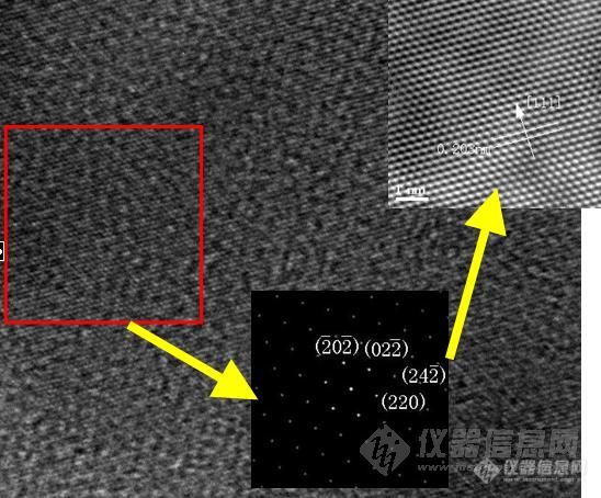
做的Ni高分辨图像,对红色选定区域进行FFT变换,Apply Mask后得到的FFT花样标定后发现各点均为(220)晶面,再对花样进行Invese FFT 后得到的二维晶格象,测量3个方向的晶面间距均为0.2034nm 对应Ni(111)晶面间距 (对原高分辨图像测量亦得到相同的晶面间距),这样高分辨的晶面间距和衍射花样的标定结果就无法自洽。由于Ni具有 FCC和 HCP 2种结构,所以对照Ni XRD结果和 Ni FCC 和HCP标准图谱发现 只能和 FCC的图谱对应,和HCP的峰位相差较远。(其中测试出的Ni只有峰位为76度的(220)方向的峰,具有高取向性)。由于对多种原因进行分析 对 HRTEM图像不能自洽的问题都得不到合理解释,现求助各位老师,同学,期待大家可以给与解答。[img]http://ng1.17img.cn/bbsfiles/images/2007/07/200707041057_57117_1843436_3.jpg[/img]
 400-860-2797
 留言咨询
留言咨询
 400-803-1678
留言咨询
400-803-1678
留言咨询
 400-803-1678
留言咨询
400-803-1678
留言咨询


 我要推广仪器
 下载APP
下载APP