

方案详情文
智能文字提取功能测试中
View Article OnlineView Journal|View Issue View Article OnlineiRSC AdvancesPaper Elemental imaging of leaves from the metalhyperaccumulating plant Noccaea caerulescensshows different spatial distribution of Ni, Zn andCdt Damien L. Callahan,*a Dominic J. Hare, David P. Bishop, Philip A. Dobleand Ute Roessner° Elemental imaging using laser ablation inductively coupled plasma mass spectrometry was performed onwhole leaves of the hyperaccumulating plant Noccaea caerulescens after treatments with either Ni, Znor Cd. These detailed elemental images reveal differences in the spatial distribution of these threeelementsacrosstheleaf andprovidennew insights innthenmetalalion homeostasis withinhyperaccumulating plants. In the Zn treated plants, Zn accumulated in the leaf tip while Mn was co-localised with Zn suggesting similar storage mechanisms for these two metals. These data show a Znconcentration difference of up to 13-fold higher in the distal part of the leaf. Also, there was nocorrelation between the S and Zn concentrations providing further evidence against S-binding ligands. Incontrast, Ni was more evenly distributed while a more heterogeneous distribution of Cd was presentwith some high levels on leaf edges, suggesting that different storage and transport mechanisms areused for the hyperaccumulation of these two metals. These results show the importance of correctsampling when carrying out subcellular localisation studies as the hyperaccumulated elements are notnecessarily homogenously distributed over the entire leaf area. The results also have great implicationsfor biotechnological applications of N. caerulescens showing that it may be possible to use themechanisms employed by N. caerulescens to increase the Zn concentration in nutrient poor cropswithout increasing the risk of accumulating other toxic elements such as Ni and Cd. Introduction A disparate group of plants found in metal rich soils worldwidecan accumulate extraordinary concentrations of metal ions ormetalloids in their aerial tissues to concentrations which wouldbe toxic to most other organisms. These plants are classified ashyperaccumulators and have developed unique mechanisms thatenable the extreme accumulation of metal ions withoutsuffering typical toxicity symptoms. At high concentrations, theselectivity of transition elements to molecules containingnitrogen, oxygen and sulphur plus their access to differentredox states result in extreme toxicity as the excess metal ionsinterfere with enzyme function and nitrogen metabolism,inhibit mitotic activities, cause oxidative damage and reduce ( "Deakin U niversity, School of Life and Environmental Sciences, C entre f or Chemistryand Biotechnology, B urwood, Geelong, Vi c toria, 3 1 25, Australia. E - mail: Damien.Callahan@deakin.edu.au ) ( Elemental Bio-imaging Fac i lity, Un i versity of Technology Sydney, Aust r alia ) ( Metabolomics A u stralia, Sc hool o f BioSciences, The Un i versity of Melbourne,Parkville, V ictoria, 3010, Australia ) ( t Electronic supplementary information (ESI) available. See DOI: 10.1039/c5ra23953b ) the uptake of other essential micronutrients.2-6 For thesereasons, the transport, distribution and storage of thesemetallic micronutrients must be actively controlled. Over-accumulation of one metal ion can disrupt the overall metalionIhomeostasis. There have beenhmany studies whichdescribed the effects of heavy metal exposure in plants, forexample, the review by Clemens contains an excellent descrip-tion of these responses with a particular focus on Cd. Most hyperaccumulators have developed a tolerance fora particular metal ion and this tolerance is typically related tothe soil substrate. The mechanisms behind this specificity andthe detoxification mechanisms are not completely understood.Part of the challenge in studying hyperaccumulators is the greatdiversity in the types of plants which have these traits since theyrange from large rainforest trees such as Pycnandra acuminata(previously Sebertia acuminata) endemic to New Caledonia tosmall species belonging to the Brassicaceae family foundthroughout Europe. For hyperaccumulation to occur the metalions in above ground tissue must be sequestered and stored insubcellular structures in order to inhibit disruption of normalmetabolic processes within the leaf. Noccaea caerulescens J & C Presl.) F. K. Mey. Brassicaceae(formerly Thlaspi caerulescens) has been identified as a modelspecies for studying hyperaccumulation.8, This species has theability to hyperaccumulate Ni, Zn and Cd. Of note is thatecotypes from different metalliferous soils throughout Europeexhibit large variations in their accumulation abilities andtolerances for Ni, Zn or Cd.10-13 An example of this is the Prayonecotype which can accumulate more than 30 000 mg kg-znwithout showing any typical phenotypical responses of heavymetal toxicity such as chlorosis or reduced growth, however, itdoes not accumulate Cd to the same amounts as otherecotypes. 2,14 Hyperaccumulating plants have considerable potential forremediation of contaminated soils through metal decontami-nation and re-establishment of vegetation cover. The potentialapplications of these plants have been recognised for some timeand is one of the key drivers for research on these plants,therefore numerous reviews and books have been written forexample the book by Chaney et al. nearly 20 years ago.5 Theyalso have great potential for biotechnological applicationsthrough the transfer of metal transport mechanisms intonutrient poor crops, known as biofortification. For review on thechallenges of biofortification see Antosiewicz et al.16 In this study, laser ablation-inductively coupled plasma-mass spectrometry (LA-ICP-MS) was used to investigate thespatial distribution of the hyperaccumulated metal ions Nit,Znand Cdas well as other key micronutrients in whole N.caerulescens leaves from plants that had been treated with thesemetal ions individually. This imaging technique rasters a laserbeam across samples and the ablated material is transported tothe ICP-MS using an Ar carrier gas. The ICP-MS then detectsionised elements based on the mass-to-charge ratio. A partic-ular strength of this technique is that multiple micronutrientsmay be measured in a single acquisition allowing the study ofmetal ion homeostasis and direct comparisons of micro-nutrient distribution in the same leaf. This technique has beenused to study a broad range of matrices from geological samplesto an array of different biological samples.17,18 Although it doesnot have the fine spatial resolution of scanning electronmicroscopy or X-ray microanalysis, LA-ICP-MS is highly sensi-tive, has a large linear dynamic range as well as the ability forisotopic analysis. For a review of in situ elemental analysis inplants see Lombi et al.19 There have been a number of studiesusing LA-ICP-MS on metal accumulation in plants, for example,it was used to coarsely map (750 um) Ni in the roots from theNi-hyperaccumulator Berkheya coddii Roessler, in leaves ofHelianthus annuus (sunflower) with a spatial resolution of 200um, leaves from Cu treated Elsholtzia splendens leaves, Zn andCu in roots of cucumber plants, boron in Poplar leaves andbiofortification and localisation studies in wheat grains.20-25However, this is the first study that has spatially mapped metalion distributions across whole leaves of a metal hyper-accumulating plant. This work reveals new information on thespatial distribution of hyperaccumulated metal ions and alsoillustrates the strength of this technique for studying micro-nutrients and metal ion homeostasis in plants. - ExperimentalPlant growth and experimental design Four different ecotypes of N. caerulescens were selected based ondifferent reported abilities to hyperaccumulate Ni, Zn and Cd,they also represent populations from geographically distinctareas and soil types.10-12,26 Seeds were germinated from pop-ulations in Bradford Dale (Derbyshire, U.K. -high capacity forZn accumulation), Prayon (Belgium - high capacity of Znaccumulation, low Cd), Les Bleynard (Southern France - accu-mulate Zn and Cd) and Basadre (North Spain on Ni richserpentine soil-expected high capacity to accumulate Ni and Zn)(Fig. 1). Seeds were germinated and grown in Jiffy pots (sphagnumpeat moss and wood pulp) in a glass house maintained ata maximum temperature of 22 °C with 14 hour day (supple-mented with artificial grow lights) and 10 hour night. Plantswere then watered with a 1/5 Hoagland's solution containingeither NiSO4(250uM),ZnSO4(500 pM) or CdSO4 (10 uM) as wellas anon-supplemented control group. These treatmentconcentrations allow normal growth of plants in relation tocontrol and were selected based on a previous laboratoryscreening trial. Plants were treated one week after germinationand were harvested after 3 months of growth. At least fiveindividual replicate plants were grown from each ecotype ineach of the four treatment regimes (control, Ni, Zn, Cd). Theamounts which plants accumulate also depend on the growthconditions and bioavailability of metals supplied. In order toconfirm the accumulation capabilities of the four selectedecotypes quantitative elemental analysis using an ICP-opticalemission spectrometer (OES) was carried out on dried leaftissues from these plants. The oldest leaf was selected forelemental imaging, while remaining leaves were immediatelyfrozen in liquid nitrogen and stored at -80°C. The leaves foriimaging were washed with deionised water (18.2 MQ) andmethanol, placed between Whatman filter paper and storedunder ambient conditions prior to imaging analysis. Minimalsample handling was undertaken in order to try and maintainmetal ion distribution. Total element concentrations The total quantitative measurements were carried out fromtriplicate biological replicates. Approximately 100 mg freshweight (FW) of the leaf tissue was taken from each plant, rinsedwith deionised water (18.2 MQ) and dried at 50 °C untila constant dry weight (DW) was observed. Dried and ground leafmaterial was accurately weighed (~15 mg) and acid digested at70 °C for 3 hours in Eppendorf tubes (2.5 mL) with concentratednitric acid (0.3 mL). The resulting clear acid digests weretransferred to 10 mLvolumetric flasks and made to volume withdeionised water. Acid digest were analysed with a Varian VistaICP-OES optical emission spectrometer (Varian Inc., Mel-bourne, Victoria,Australia) with the following settings: power 1kW, plasma flow 15 L min-, auxiliary flow 1.5 L minandnebuliser flow 0.9 L min-1. Instrument data were evaluatedusing Vista Pro ICP expert 4.1.0. The instrument was calibrated Fig.1(Left) Map showing the origin of seeds for each ecotype; (right) different ecotypes after three months growing in Jiffy Netted Pots (controlgroup) showing clear morphological differences. with standard solutions of Al, As, B, Cd, Cr, Cu, Fe, Hg, K, Pb,Co, Mn, Na, Ni, P, Se, V, Zn ranging between 5 mg L-and 100mg L-. These solutions were prepared by appropriate dilutionswith deionised water of the ICP mixed element stock standard(AM3, Choice Analytical, Australia) and B, K, Na, P (ChoiceAnalytical, Australia) stocks. Leaf sections were also made to quantitatively confirm thespatial distributions found by elemental imaging. The leavesthat were snap frozen and stored were rinsed with de-ionisedwater then immediately sectioned into thirds (tip, middle,base). Each section was then placed into a separate pre-weighed2 mL Eppendorf tube. This approach removes the chance for re-distribution of metal ions within the leaf tissues. Fresh weightsand dry weights were recorded. The samples were then aciddigested as above and elements quantified by ICP-MS (Perki-nElmer NexION 350-X). Laser ablation ICP-MS imaging Samples were analysed using an Agilent 7500 ICP-MS (AgilentTechnologies, Victoria, Australia) coupled to a New WaveTechnologies UP-213 laser ablation system (Kennelec Technol-ogies, Victoria, Australia). The laser ablation system containeda Nd:YAG laser and a large format laser ablation cell of 15.2 ×15.2 cm. The cell employed a roving sample cup which movedthe stage in x-y-z directions while the laser beam positionremained fixed. Tygon tubing (3 mm i.d.) transferred ablatedions using a stream of argon to the ICP-MS. The ICP-MS wastuned prior to each analysis using the NIST standard referencematerial 612,trace elements in glass. The following settingswere used for the ICP-MS: RF power 1250 W, plasma gas flowrate 15 L min-, measured mass to charge ratios (m/z): C (m/z13), Cd (m/z 111), Cu (m/z 63), Fe (m/z 57), K (m/z 39), Mg (m/z24), Mn (m/z 55), Mo (m/z 95), Na (m/z23), Ni(m/z 60), P (m/z 31),S (m/z 34), Zn (m/z 66). The laser was operated at 213 nm,repetition frequency 20 Hz, laser energy 30% power, resolution80 um, scan rate 99 um s-1. Approximately 150 lines per leafwere analysed producing a total run time of 15 hours per leaf.Leaves were ablated from the top leaf surface. Each scan line produces a (.csv) file, which was collated usinga Visual Basic macro (Microsoft) and imported into ENVI (Exe-lis, Boulder, Co., USA) for image analysis." Results and discussion Total element concentration Correlating to previous studies, the unique ecotypes accumu-lated different concentrations of each metal ion (Fig.2) eventhough they were grown side by side and treated with the samesolutions. The variation in accumulation capabilities of thedifferent ecotypes provides a rich area for research but was notthe main focus of this study. The Prayon ecotype accumulatedapproximately twice the concentration of Zn (7900 mgkgdwin comparison to the next highest ecotype from Le Bleynard inSouthern France (4020 mg kg-dw). Although Prayon plantsaccumulated the highest Zn concentrations they accumulatedapproximately 10-fold less of the other two metal treatments, Niand Cd. This low accumulation factor for the Prayon populationhas been observed previously.26 The Puente Basadre ecotypeaccumulated the highest Cd concentration (960 mg kg-dw)while the Bradford Dale and Le Blaynard had the highest Niconcentrations (740 mg kg , 720 mg kgrespectively). As ex-pected all treated plants had significantly more of the addedmetal ion then the non-treated controls (ESI Fig. S1t). The Fig. 2Total Ni, Zn and Cd elemental concentrations in leaf tissuesfrom treated plants of the four different ecotypes grown. Bars aregrouped by the three treatments either CdSO4 (10 uM; black), NiSO4(250 pM; red), ZnSO4 (500 uM; green). Control plant values omittedfrom this graph due to comparatively low concentrations of theseelements. Full elemental values in Online Resource 1. Puente Basadre ecotype was selected for subsequent elementalimages as it contained the high levels of all three treated metalions. A sample of the Prayon ecotype was also imaged todetermine if there were any differences between ecotypes withregards to elemental distributions. Elemental images N. caerulescens has the ability to accumulate up to 3% Zn in itsdry mass and therefore could exceed the maximum signalintensity of the instrumentation. This would provide a non-linear response to Zn due to saturation of the detector signal.To determine the maximum concentration range of the LA-ICP-MS system, dried,ground tissues from hydroponically grown N.caerulescens plants grown in a previous study were pressed intoa disk.28 These represented tissue concentrations at 300,11 000,15 000 and 20 000 mg kg-leaf dry mass.8 The ICP-MS signalwas saturated between the 11 000-15 000 mgkg-samples. Thesamples analysed in this study were well below this thresholdand therefore represent images within the linear dynamic rangeof the instrument. Images based on the elemental intensity were produced foreach measured element and are represented here either nor-malised to the 13c isotope or plotted based on the raw signalintensity. The fine resolution of these images show obviousfeatures in the leaves, in particular leaf venation (Fig. 3-5). Astriking accumulation pattern was observed from the Zn-treatedplants (Fig. 3). Significantly higher concentrations of Zn wereclearly identified in the leaf tip. A Zn concentration gradient ispresent from the leaf tip to approximately half way down thelamina. The relative differences in elemental concentrations 0 0.125 >0.2560Ni:13C signal intensity 0 0.0075 >0.015 1Cd:13C signal intensity Fig. 4Ni (top) and Cd (bottom) images from the different treatmentsfrom the Puente Basadre population. The Ni elemental image showsa relatively homogenous distribution, while Cd shows a moreheterogeneous distribution with some increased Cd concentration onthe leaf edges.Note in the signal intensity from the plants not treatedwith the corresponding metal are outlined due to the low relativeintensity. Non-normalised images are in ESl.t were determined using the mean signal intensity from keyspatial regions of the leaf. For the Zn image, this representsa 13-fold higher concentration of Zn at the leaf tip compared tothe base of the leaf (Fig. 3). The bottom half of the lamina(closest to the petiole) contained a relatively homogenous Znconcentration. The Zn-concentration supplied to the plants was Fig.3Elemental images from the Puente Basadre population.Top, Zn heatmap showing distribution in leaves from a Zn treated plant and controlplant (outlined due to low relative signal). Zn intensity was normalised using the 13C signal. Bottom panel showing a similar distribution for Mn. Fig.5 Etlemental images based on the raw signal intensity of potassium, phosphorous, magnesium, sulfur, carbon, and copper from Zn treatedplant showing a homogenous distribution of these macro elements. The intensity values of these images illustrated on the bar to the right of eachimage. Not some contamination of leaf surface can be seen in the copper image. All images available in ESl 4.+ constant throughout the 3 month growth period and thereforethe plant is actively concentrating Zn at the leaf tip. It is alsoclear from these images that Zn was lower in concentration inthe vascular tissues, where the leaf veins are darker and there-fore lower in Zn-concentration. The Zn treated Prayon ecotypewas also imaged had the same Zn-accumulation pattern as thePuente Basadre ecotype suggesting similar storage mechanismsfor Zn (ESI Fig.S2t). Examination of other images showed a veryclear co-localisation of Mn and Zn (Fig. 3). From previous workin our laboratory, a clear reduction in Mn was observed whenthe plant Zn concentration exceeded 10 000 mg kg(ESIFig. S3t).28 This suggests that Zn and Mn have similar uptakemechanisms and also explains why the Mn concentrationdecreases when N. caerulescens accumulates extremely highconcentrations of Zn. No other elements showed the samestriking co-localisation patterns in the Zn-treated plants.Sulphur showed some increase in intensity (2-fold difference) atthe leaf tip but not to the same extent of Zn providing furtherevidence against S-binding ligands as part of storage in N.caerulescens.2The Zn image of the non-treated control leaf onthe same scale ofthe treated plants is hardly discernible (Fig.3). Fig.6Zoomed in image of Zn (top) and Cd (bottom). Higher Znconcentrations are located outside the vascular tissue in contrast toCd. Images are produced on the raw signal intensity. However, in the leaf from a control plant, which has a 3.5-foldlower total leaf Zn concentration, a similar accumulationpattern was observed with higher concentrations in the leaf tip(2-fold). No other accumulation patterns were observed for theother elements in the control plants (ESI Fig. S4t). From the Feand Cu images it appears that some leaf contaminationoccurred which was not removed by washing. This most likelyoccurred during the supply of nutrient solutions to the plantscausingthe precipitation of insoluble Fe and Cu salts on the leafsurface (Fig. 5, ESI Fig S4t). However it can still be observed inthe Fe and Cu images that concentrations of these elements arenot effected by high localised concentrations of Zn. The samehomogenous distribution is also observed in leaves from plantstreated with Ni and Cd. As expected the elemental images from the Ni and Cd treatedplants show a huge contrast in signal intensities relative to thenon-treated plants (Fig. 4). The elemental images in the Ni andCd treated plants did not show any distinet accumulationpatterns. Ni was relatively evenly distributed across the wholeleaf with lower concentrations in leaf vascular tissue (Fig. 4). Noclear pattern could be observed for Cd distribution apart fromhaving higher concentrations in leaf vascular tissue and someof the leaf edges (Fig. 5 and 6). The more heterogeneous Cddistribution is in contrast to Zn and Ni. These images reveal thateven though multiple elements are hyperaccumulated by N.caerulescens, different storage strategies are used for Ni, Zn andCd. It is evident that in N. caerulescens Zn accumulation beginsat the distal part of the lamina. In contrast, there is no regionalconcentration for Ni. A closer examination of the Cd and Cuimages show higher concentrations in the vascular tissues ofthe lamina in comparison to surrounding tissue which alsocontrasts to Zn and Ni. This is most apparent in the zoomed inelemental images of Zn versus Cd (Fig.6). As discussed abovethe elemental imaging technique used here cannot produceimages which would enable conclusions to be made at thesubcellular level. Molecular studies have shown that Zn istransported symplasticallybyy various highly expressedmembrane transporters such as the HMA4 (Heavy Metal ATPase4) and ZIP (Zn regulated transporter, iron-regulated transporter- Table1LQuantitative confirmation of elemental distributions found byelemental imaging. Results are from plants treated with either Ni, Zn orCd. The leaves were sectioned into thirds; base, middle and tip, dried,acid digested and analysed by ICP-MS Base Mid Tip Zn (mg kg) dw Zn-1 420 1400 1700 Zn-2 239 600 1200 Zn-3 150 640 1600 Zn-4 1500 3700 6500 Ni (mg kg) dw Ni-1 370 550 630 Ni-2 590 700 960 Ni-3 410 1900 1300 Cd(mg kg) dw Cd-1 210 370 340 Cd-2 150 300 380 Cd-3 190 220 230 related protein) gene families.32,33 With regards to Cd there is nosolid evidence for apoplastic localisation of Cd in leaves. A veryearly study which claimed apoplastic storage was most likelydue to sample preparation artefact due to the chemical fixationtechnique used.3 It was subsequently shown that Cd is activelytaken up into the epidermal storage cells.31,34 The distribution of macro elements such as Na, K, Mg, S andFe show a homogenous distribution throughout the leaves in alltreatments. This shows that the concentrations of these keyelements are not affected by the presence of high concentra-tions of the hyperaccumulated metal ion (Fig. 5). As the imaging data showed differences elemental spatialdistributions,quantitative measurementswere madee toconfirm that the patterns observed were not an artefact ofsample preparation. These quantitative measurements werealso carried out in triplicate to provide replication of theseobservations. The leaves were sectioned into thirds: tip, middleand base, oven dried, acid digested then total elementalconcentrationsdeterminedbysolution ICP-MS.Thesemeasurements confirm the elemental imaging data (Table 1)with 4-10 fold higher concentrations of Zn in the leaf tip andmore homogenous distributions of Ni and Cd. Only a few published studies to date have examined themacroleafspatial distribution(of elements in hyper-accumulators. One study analysed leaf cross sections usingmicro-PIXE and found a homogenous distribution of Ni indifferent cell types in the Australian Ni hyperaccumulatorStackhousia tryonii, suggesting that a similar accumulationpattern was observed to this study.35 The radioactive 109Cdisotope was used previously to image Cd compartmentation atthe leaf scale in the hyperaccumulators N. caerulescens, Arabi-dopsis halleri and willow leaves.36-38 These studies agree with theresults described here. In the three species referenced theauthors showed a heterogeneous Cd distribution in the olderleaves, in particular, high concentrations in vascular tissues and leaf edges. The paper by Cosio et al. (2005) is of particularrelevance to this work. The Cd autoradiography was carried outon two contrasting ecotypes of N. caerulescens. This work clearlyshowed Cd localised at leaf edges as well as points of higherconcentration spread out over the whole leaf surface. X-raymicroanalysis of the Cd cellular distribution showed that Cdwas stored in less metabolically active areas of leaf cells.Unfortunately this study did not carry out a similar analysis forZn. An early study comparing Cd and Zn uptake also showeddifferent distribution patterns for these metals based on leafage.3 The study by Perronnet et al. (2003) showed that Cdconcentrations remained relatively constant with increasingbiomass while Zn concentrations had a small decrease withtime. It has also been shown that Cd is not re-distributed fromold to young organs during leaf senescence suggesting that Cdions are fixed in place once accumulated.4 This is in contrast toZn which is actively take up and concentrated in leaf tissues byN. caerulescens.41 Other studies have shown that the Cd co-ordination environment changes with tissue age. For example,tissue age differences in the concentration and the co-ordination environment of Cd were observed N. caerulescensleaf tissue using X-ray absorption spectroscopy.2 The determination of the spatial distribution of elements isimportant for understanding how hyperaccumulators transportand store the large amounts of metals in a way that does notinterfere with essential metabolic processes and maintainselemental homeostasis. The findings here also have implica-tions for tissue sampling. For example, a recent study focussedon trace elements in hyperaccumulators from New Caledonia, itwas shown concentrations of Mn increase in the older leaves ofthe hyperaccumulator Grevillea meisneri.43 These findingsillustrate the importance of correct sampling and the consid-eration of leaf age when determining the ability and themechanisms of plants to accumulate trace elements. Most research on metal ion distribution in hyperaccumulatorshas focused on cellular and sub-cellular distributions as opposedto whole tissues as was carried out in this research. For a reviewon elemental distribution in plants see Zhao et al. 2014.44 Therehave been some conflicting reports of the cellular metal iondistribution in N. caerulescens and other closely related speciesusing X-ray microanalysis techniques. For example, organelleisolation found that 65-70% of the total leaf Cd and Zn waslocated in vacuoles of mesophyll tissues in N. caerulescens.However, other studies have shown Zn storage in the epidermiscells instead of the metabolically active mesophyll layer.46-48 Theconflict here may be due to different sample preparation tech-niques. The paper by Ma et al. (2005) removed the epidermallayer. This may have resulted in the rupture of epidermal cellswith the cell contents transferring to the mesophyll layer. Here we have shown the importance of selecting the correctareas within the leaf to study subcellular localisation and to firstconfirm if there is a difference on the spatial distribution ofelements. A single leaf cross section may not represent the samedistribution across the whole leaf. The spatial resolution of the LA-ICP-MS does not allow imaging of subcellular structures,howeverit is highly sensitive and whole leaf elemental images still provideuseful information on the metal-ion homeostasis in plants. The large variation observed in the total elemental concentra-tions of the four ecotypes illustrates the rich diversity present inthe N. caerulescens hyperaccumulator. The use of LA-ICP-MS hasallowed multiple elemental images to be developed without theuse of radioactive labelling and with minimal sample prepara-tion, however, the analysis itself is destructive. This technique ishighly sensitive and has a large dynamic range that enablesmeasurements of trace elements as well as hyperaccumulatedmetals in plants. In this study it was revealed that Zn accumu-lates distal area within the leaf and co-localises with Mn and tosome degree S. No other elements showed co-localisation withZn. In contrast to Zn, Ni is more homogenously distributedacross the leaf while a more un-even distribution of Cd wasfound, with clearly higher concentrations in vascular tissues.These results show the importance of correct sampling whenstudying hyperaccumulators as the hyperaccumulated elementsmay not be distributed evenly across a leaf and thereforesubcellular studies must take this into account. Finally, as Zn is stored differently to Ni, and Cd it may bepossible to utilise the selectivity of the Zn transporters used byN. caerulescens in future biofortification studies without the riskof increasing Ni and Cd concentrations. Acknowledgements This research was supported by funding from Deakin Universityand the central research grants scheme (CRGS) and an earlycareer researcher grant from The University of Melbourne. DLCallahan would like to thank Vasalisa Pendan for help withplant growth. The authors would also like to thank Prof. AlanBaker for provision of the N. caerulescens seed bank. References ( 1 U. Kramer, Annu. R e v. P l ant Biol., 2010, 61,5 1 7-534. ) ( 2 C. Anderson, A . Deram, D. Petit, R. Brooks, R. Stewart and R. S imcock, i n T r ace El e ments in S oil: Bi o availability, Flux, and T ransfer, ed. I . K. Iskandar and M. B. Kirkham, CRC Press, Boca Raton, 2001, ch. 4, pp.63-76. ) ( 3 C. Chen, D. H uang and J. Liu, Clean:Soil, Air, Water, 2009,37, 304-313. ) ( 4 E. Gajewska, M. W ielanek, K. Bergier and M . Sklodowska,Acta Physiol. Plant. , 2009 , 31 , 1291-1300. ) ( 5 K . V. Madhava Ra o and T. V. S. Sresty, Plant Sci., 2000, 157,113-128. ) ( 6 J. Molas, Environ. Exp. Bot., 2002, 47, 115-126. ) ( 7 S.Clemens, Biochimie, 2006,88,1707-1719. ) ( .8 A. G. L . Assuncao, H. Schat and M. G. M. Aarts, New Phytol., 2003, 159,351-360. ) ( 9 M. J. Milner and L . V. Kochian, Ann. B ot., 2008, 102,3-13. ) ( 10 A. Baker, R. Reeves and A. Hajar, New Phytol., 1994, 127, 61- 68. ) 11 R. D. Reeves, C. Schwartz,J. L. Morel and J. Edmondson, Int.J. Phytorem., 2001,3,145-172. 12 E. Lombi, F. J. Zhao, S. J. Dunham and S. P. McGrath, NewPhytol., 2000, 145, 11-20. 13 A. G. L. Assuncao, W. M. Bookum, H. J. M. Nelissen,R. Vooijs, H. Schat and W. H. O. Ernst, New Phytol., 2003,159,411-419. 14 S. L. Brown, R. L. Chaney,J. S. Angle and A. J. M. Baker, SoilSci. Soc. Am. J.,1995,59,125. 15 R. L. Chaney, M. Malik, Y. M. Li, S. L. Brown, E. P. Brewer,J. S. Angle and A. J. Baker, Curr. Opin. Biotechnol., 1997, 8,279-284. ( 16 D.M. Antosiewicz, A. Barabasz and O. Siemianowski, FrontPlant Sci., 2014, 5, 80. ) ( 17J. S . Becker, M. Zoriy, A. Matusch, B. Wu, D . Salber, C. Palmand J. S. Becker, Mass Spectrom. Rev.,2010, 2 9, 156-175. ) 18 D.Pozebon.,G.L. Scheffler. V. L.DresslerandM. A. G. Nunes,J. Anal. At. Spectrom.,2014,29,2204-2228. ( 19 E. Lombi, K. G. Scheckel a nd I. M. K empson, Environ. E x p. Bot.,2011,72,3-17. ) ( 20 A. B. Moradi, S. Swoboda, B . R obinson, T. P rohaska,A. Kaestner, S . E. Oswald, W. W. Wenzel and R. Schulin,Environ. E xp. B ot., 2010, 69, 24-31. ) ( 21 B . Wu, M. Zoriy, Y. Chen and J. S. Becker, Talanta, 2009,78, 132-137. ) ( 22 J . Shi, M. A. Gras and W. K. Silk, Planta, 2009, 229,945-954. ) ( 23 R. Rees, B. H. [. Robinson, M. l .. Menon, E. .I Lehmann, M. S . G unthardt-Goerg a n d R. Sch u lin, Environ. Sci.Technol.,2011,45,10538-10543. ) ( 24 I. Cakmak, M . Kalayci, Y . K aya, A . A . Torun, N. Aydin,Y. Wang, Z. A risoy, H. Erdem, A . Y azici, O. Gokmen, L. Ozturk and W . J. H o rst, J. Agric. F ood Chem., 2010, 58,9092-9102. ) ( 25 B . Wu, F. Andersch, W. Weschke, H. Weber and J. S. Becker,Metallomics, 2 013, 5, 1276-1284. ) ( 26 N. Roosens, N. Verbruggen, P. Meerts, P. Ximenez-Embunand J. A. C. Smith, Plant, C ell Environ., 2003, 26,1657-1672. ) ( 27 D. Hare, B . R eedy, R. G r imm, S. W il k ins, I. Vol i takis,J. L. George, R . A . C herny, A. I. B u sh, D. I. Finkelstein an d P.Doble, Metallomics, 2009, 1, 53-58. ) ( 28 S . Foroughi, A. J. M . Baker, U . R o essner, A. A. T. Jo h nson, A. Bacic and D . L. Callahan, Metallomics, 2014,6,1671-168 2 . ) ( 29 V. H. Hassinen, M. Tuomainen, S . P e raniemi, H. Schat,S. O. K arenlampi and A. I. Te r vahauta, J. Ex p . Bo t ., 2009, 60, 187-196. ) ( 30 M. D . Vázquez, J . B a rcelo, C . P o schenrieder, J. M adico,P. Hatton, A. J. M. B aker a n d G. H. Co p e,J. Plant Physiol., 1992,140,350-355. ) ( 31 M. W ojcik, J. Vangronsveld, J. D 'Haen a n d A. Tukiendorf, Environ. Exp. Bot. , 2005,53,163-171. ) ( 32 A . Assuncao, P. Martins, S. de Folter, R. Vooijs, H.Schat andM. A arts, P lant, Cell Environ., 2001,24,2 1 7-226. ) ( 33 N. S. Pence, P. B. Larsen, S. D. Ebbs, D. L. L etham,M. M . Lasat, D. F. G arvin, D. Eide and L. V . K o chian, Proc.Natl. Acad. Sci. U. S. A., 2000, 97, 4956-4960. ) ( 34 B . Leitenmaier and H. Kupper, Plant, Ce l l Environ., 2011, 34, 208-219. ) 35 N. P. Bhatia, K. B. Walsh, I. Orlic, R. Siegele, N. Ashwath andA. J. M. Baker, Funct. Plant Biol., 2004, 31, 1061-1074. ( 36 C . Cosio, L. DeSantis,B. F r ey,S. Diallo a n d C. Keller, J . E xp. Bot.,2005,56,765-775. ) 37 C. Cosio, P. Vollenweider and C. Keller, Environ. Exp. Bot.,2006,58,64-74. 38 S. Huguet, V. Bert, A. Laboudigue, V. Barthes, M.-P. Isaure,I. Llorens, H. Schat and G. Sarret, Environ. Exp. Bot., 2012,82, 54-65. ( 39 K . P erronnet, C . S c hwartz and J. Mo r el, Plant Soil, 20 0 3, 249, 19-25. ) 40 L. Lovy,D. Latt and T. Sterckeman, Plant Soil, 2013, 362,345-354. 41 S. N. Whiting,J. R. Leake,S. P. McGrath and A. J. M. Baker,New Phytol.,2000, 145,199-210. ( 42 H. Kupper, A. M ijovilovich, W. Meyer-Klaucke and P. M.H. K r oneck, Plant Physiol., 2004,134, 7 48 -757. ) 43 G. Losfeld, L. L'Huillier, B. Fogliani, S. Coy, C. Grison andT. Jaffre, Environ. Sci. Pollut. Res., 2015,22,5620-5632. 44 F.-J. Zhao, K. L. Moore, E. Lombi and Y.-G. Zhu, Trends PlantSci.,2014,19,183-192. ( 45 J. F. Ma, D. Ueno, F.-J. Zhao and S. P. McGrath,Planta, 2005, 220,731-736. ) 46 U. Kramer, I. J. Pickering, R. C. Prince, I. Raskin andD. E. Salt, Plant Physiol., 2000, 122, 1343-1353. ( 47 H . K iipper, F . J ie Zhao and S. P. M c Grath, Plant Phy s iol.,1999, 119,305-312. ) ( 48 K . Vogel-Mikus, J. Simcic, P. Pelicon , M. Budnar, P. Kump,M. N ecemer, J . Mesjasz-Przybylowicz, W. J. P r zybylowiczand M . Regvar, Plant, Cell Environ., 2008,31,1484-1496. ) RSC Adv., , his journal is O The Royal Society of Chemistry 使用激光剥蚀电感耦合等离子体质谱法,对处理后的整个超富集蓝藻植物叶中的镍,锌或镉进行成像。这些详细的元素图像展示了这三个要素在蓝藻叶中的空间分布差异,为研究超富集植物内部中元素平衡研究提供了思路。在锌处理的植株中,锌积累在叶尖,而锰与锌共渗,表明这两种金属的储存机制相似。这些数据显示,在叶片的远端,锌的浓度差异高达13倍。此外,硫和锌浓度之间也没有相关性,这进一步证明了硫结合配体是不对的。而镍的分布较为均匀,镉的分布较为不均一,在叶片边缘有较高的含量,说明这两种金属的超富集利用了不同的储运机制。这些结果表明,在进行亚细胞定位研究时,正确采样的重要性,因为超积累元素不一定均匀分布在整个叶片区域。这一结果也对豆科植物的生物技术应用具有重要意义,表明豆科植物在不增加积累镍、镉等其他有毒元素的情况下,可以利用豆科植物所使用的机制提高营养不良作物中锌的浓度。
关闭-
1/8
-
2/8
还剩6页未读,是否继续阅读?
继续免费阅读全文产品配置单
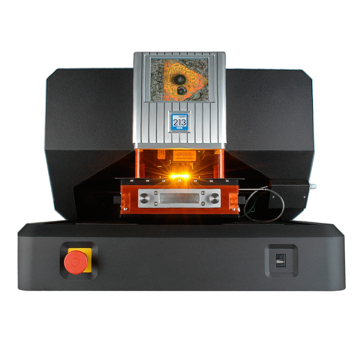
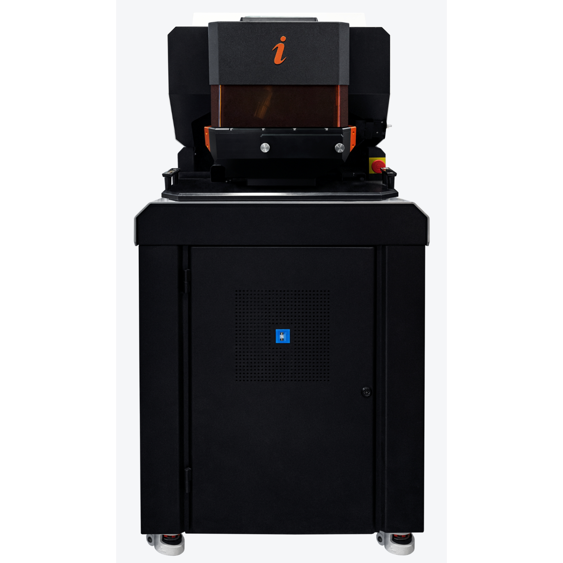
上海凯来仪器有限公司为您提供《超富集植物蓝藻叶片中Ni、Zn和Cd的空间分布检测方案(激光剥蚀进样)》,该方案主要用于其他中Ni、Zn和Cd的空间分布检测,参考标准《暂无》,《超富集植物蓝藻叶片中Ni、Zn和Cd的空间分布检测方案(激光剥蚀进样)》用到的仪器有ESL213 灵活的激光剥蚀系统。
我要纠错
推荐专场
相关方案
 咨询
咨询