

 仪器对比
仪器对比


 关注
关注
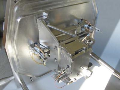
3VIEW™
————立体表面成像新技术,3D成像技术的革命
3 VIEW™ 专门设计了一种适用性极广泛的3D成像技术。它可以在低真空或环境场发射扫描电镜中实现对样品的原位观察,自动获取超精细3D结构。3VIEW不仅对立体表面的成像建立序列关系,并且在序列之间建立对应。这样的优点在于可以简单地将图像输出到第三方软件的3D成像系统,而不需要对图像进行繁琐的后处理。适合的研究领域有:神经系统科学,干细胞, 胚胎学, 病理学, 植物学,组织培养.等等
Block face imaging in the SEM
3View™ uses a specially designed ultra-microtome operating in situ within a variable pressure, Field Emission SEM, and allows automated acquisition of 3D ultrastructure by sequentially imaging the freshly cut, resin embedded block face.
Perfect image stack alignment
Due to the technique of serially imaging the block face rather than individual sections, and because of the stability of 3View's stage, there is excellent registration between all images within an image stack. This allows simple exporting of the image files for 3-D rendering by 3rd party software, without the need for tedious post-processing.
High resolution results
3View’s unique design allows a relatively short SEM working distance to stay exactly constant throughout a cutting sequence. Furthermore, a finely focused electron probe and small interaction volume enables a spatial resolution over 1 order of magnitude superior to that of confocal microscopy, and approaching the nanometer scale. As there is no depth probing, this 3D resolution is maintained throughout the sectioning depth.
3View data of retina with extra-cellular horseradish peroxidase (HRP). Horstmann, Euler, Hausselt, Briggman, Denk; MPI for Medical Research Heidelberg are kindly acknowledged for their collaboration and specimen preparation.
Depending on the specimen and its preparation, 30nm thickness slice removal can be obtained. Similar fine depth resolution in the image contrast mechanism can be achieved with optimised injection conditions and a very high sensitivity BSE detector.
Stability for large ‘image stacks’
No wasted time
Imaging over a significant volume
Unattended operation
Easy to use
One software platform
Specimen preparation, image contrast and resolution
Specimen preparation and the image contrast mechanism are based on heavy metal staining, fixing and resin-embedding, a technique well established in the field of TEM of thin sections.
Specimen blocks are ideally pre-trimmed and faced before being transferred to the in situ 3View system.
Back-Scattered Electron (BSE) imaging is used to show ultra-structural biological detail according to the local density of the stain heavy metal atoms. A stronger BSE signal is associated with denser regions of high atomic weight. As TEM microscopy is well established in this field, 3View provides images of similar contrast, (which is simply the reverse polarity of a standard SEM BSE signal).
A Field Emission SEM is required for best results as this enables high spatial resolution imaging with the required long term stability. Variable pressure mode is required to avoid resin sample charging. A small spot size helps maximize spatial resolution, but since the BSE signal is being measured, the ultimate spatial resolution will depend on the specimen preparation, and column conditions required for adequate contrast and signal to noise. For maximum 3D spatial resolution, and to avoid charging, very low injection conditions are required and an optimised BSE detector is essential for this application.
技术参数:
System Description
High-stability in-situ ultra-microtome, stage and imaging system, to allow Serial Block Face Scanning Electron Microscopy within a Variable Pressure Field Emission SEM*.
1. the knife is aligned with the block face and ready to cut
2. the knife is moving and shaving the surface of the block removing 50nm resin
3. the knife has shaved the surface and is retracting to its original position
4. the surface of the block is scanned and the backscattered electrons generate the image
Includes:
High precision in-situ ultra-microtome, capable of cutting slices of < 50nm
Diamond knife
High stability x-y- stage (movements sufficient to cover 1mm x 1mm specimen block)
SEM stage door and low noise electrical feedthroughs
System electronics for software control of microtome and stage movement, and DigiScan IITM for additional microtome control and image acquisition
Optical microscope, bench top alignment stand, stage door mounting stand, lighting and live movie camera to aid setup and coarse specimen approach
Optional : Special BSE detector, optimised for low kV imaging
3View software features
Setup monitoring
Video streaming from optical microscope
主要特点:
获得高精确的原位图像,步长<50nm
钻石切割刀
高稳定性的,可X,Y双轴调节的样品台(1mmX1mm)
快速的图像读取
高的空间分辨率,大视野
操作简单方便
3View software features
Setup monitoring
Video streaming from optical microscope
DigitalMicrographTM Control and Acquisition
Control of knife and specimen advance / retract parameters
SEM column communication for magnification, focus, astigmatism, beam blanking
DigiScan IITM digital beam control for imaging with wide choice of pixel density, aspect ratio, pixel dwell time, persistence for search and preview, multi frame integrate for record mode and simultaneous imaging inputs
Control of stage position, including point and go feature with automatic backlash removal
Column communication for control / knowledge of external scan control, magnification, focus, astigmatism, beam blanking
Automatic 3D acquisition of single or multiple slices at different locations and/or magnifications
Image monitoring to initiate debris cleaning or other thresholding actions.
Reviewing of live 3D stacks during acquisitions
Configurable auto-survey routines
Post Processing and Exporting Tools
3D volume rebinning tools including creation of 3D “thumbnails”
Slice viewer and slice movie player, applicable to 3D file in RAM
Browser tool, applicable to files larger than RAM capability
Filtering and export tools for use with 3rd party 3D software routines


相关产品

