
第1楼2005/04/02
How it works
Atoms of different elements absorb characteristic
wavelengths of light. Analysing a sample to see if it
contains a particular element means using light from
that element. For example with lead, a lamp
containing lead emits light from excited lead atoms
that produce the right mix of wavelengths to be
absorbed by any lead atoms from the sample. In
AAS, the sample is atomised – ie converted into
ground state free atoms in the vapour state – and a
beam of electromagnetic radiation emitted from
excited lead atoms is passed through the vaporised
sample. Some of the radiation is absorbed by the lead
atoms in the sample. The greater the number ofatoms there is in the vapour, the more radiation is
absorbed. The amount of light absorbed is
proportional to the number of lead atoms. A
calibration curve is constructed by running several
samples of known lead concentration under the same
conditions as the unknown. The amount the
standard absorbs is compared with the calibration
curve and this enables the calculation of the lead
concentration in the unknown sample.
Consequently an atomic absorption spectrometer
needs the following three components: a light source;
a sample cell to produce gaseous atoms; and a means
of measuring the specific light absorbed.
第2楼2005/04/02
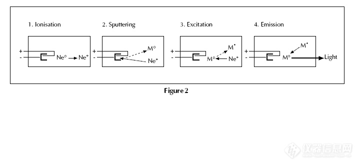
The light source
The common source of light is a ‘hollow cathode
lamp’ (Fig. 1). This contains a tungsten anode and a
cylindrical hollow cathode made of the element to be
determined. These are sealed in a glass tube filled
with an inert gas – eg neon or argon – at a pressure o
between 1 Nm–2 and 5 Nm–2. The ionisation of some
gas atoms occurs by applying a potential difference of
about 300–400 V between the anode and the
cathode. These gaseous ions bombard the cathode
and eject metal atoms from the cathode in a process
called sputtering. Some sputtered atoms are in
excited states and emit radiation characteristic of the
metal as they fall back to the ground state – eg
Pb* → Pb + h (Fig. 2). The shape of the cathode
concentrates the radiation into a beam which passes
through a quartz window, and the shape of the lamp
is such that most of the sputtered atoms are
redeposited on the cathode.
第4楼2005/04/02
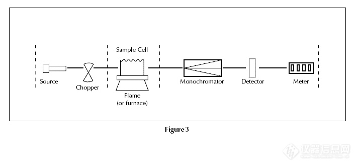
The optical system and detector
A monochromator is used to select the specific
wavelength of light – ie spectral line – which is
absorbed by the sample, and to exclude other
wavelengths. The selection of the specific light allows
the determination of the selected element in the
presence of others. The light selected by the
monochromator is directed onto a detector that is
typically a photomultiplier tube. This produces an
electrical signal proportional to the light intensity
(Fig. 3).
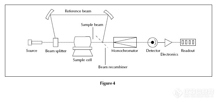
Double beam spectrometers
Modern spectrometers incorporate a beam splitter so
that one part of the beam passes through the sample
cell and the other is the reference (Fig. 4). The
intensity of the light source may not stay constant
during an analysis. If only a single beam is used to pass
through the atom cell, a blank reading containing no
analyte (substance to be analysed) would have to be
taken first, setting the absorbance at zero. If the
intensity of the source changes by the time the
sample is put in place, the measurement will be
inaccurate. In the double beam instrument there is a
constant monitoring between the reference beam and
the light source. To ensure that the spectrum does not
suffer from loss of sensitivity, the beam splitter is
designed so that as high a proportion as possible of
the energy of the lamp beam passes through the
sample.
第6楼2005/04/02
Atomisation of the sample
Two systems are commonly used to produce atoms
from the sample. Aspiration involves sucking a
solution of the sample into a flame; and
electrothermal atomisation is where a drop of sample
is placed into a graphite tube that is then heated
electrically.
Some instruments have both atomisation systems
but share one set of lamps. Once the appropriate lamp
has been selected, it is pointed towards one or other
atomisation system.
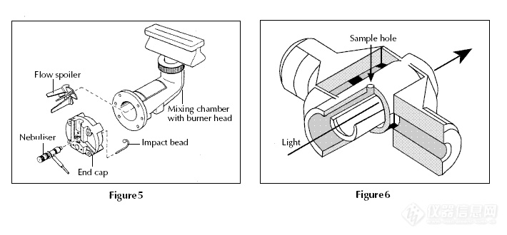
Flame aspiration
Figure 5 shows a typical burner and spray chamber.
Ethyne/air (giving a flame with a temperature of
2200–2400 °C) or ethyne/dinitrogen oxide (2600–
2800 °C) are often used. A flexible capillary tube
connects the solution to the nebuliser. At the tip of
the capillary, the solution is ‘nebulised’ – ie broken
into small drops. The larger drops fall out and drain
off while smaller ones vaporise in the flame. Only
ca 1% of the sample is nebulised.
第8楼2005/04/02
Electrothermal atomisation
Figure 6 shows a hollow graphite tube with a platform.
25 μl of sample (ca 1/100th of a raindrop) is placed
through the sample hole and onto the platform from
an automated micropipette and sample changer. The
tube is heated electrically by passing a current
through it in a pre-programmed series of steps. The
details will vary with the sample but typically they
might be 30–40 seconds at 150 °C to evaporate the
solvent, 30 seconds at 600 °C to drive off any volatile
organic material and char the sample to ash, and with
a very fast heating rate (ca 1500 °C s-1) to 2000–
2500 °C for 5–10 seconds to vaporise and atomise
elements (including the element being analysed).
Finally heating the tube to a still higher temperature
– ca 2700 °C – cleans it ready for the next sample.
During this heating cycle the graphite tube is flushed
with argon gas to prevent the tube burning away. In
electrothermal atomisation almost 100% of the
sample is atomised. This makes the technique much
more sensitive than flame AAS.
第9楼2005/04/02
Sample preparation
Sample preparation is often simple, and the chemical
form of the element is usually unimportant. This is
because atomisation converts the sample into free
atoms irrespective of its initial state. The sample is
weighed and made into a solution by suitable
dilution. Elements in biological fluids such as urine
and blood are often measured simply after a dilution
of the original sample. Figure 7 shows a flame atomic
absorption spectrometer with an autosampler and
flow injection accessory.
When making reference solutions of the element
under analysis, for calibration, the chemical
environment of the sample should be matched as
closely as possible – ie the analyte should be in the
same compound and the same solvent. Teflon
containers may be used when analysing very dilute
solutions because elements such as lead are sometimes
leached out of glass vessels and can affect the results.
第10楼2005/04/02
Background absorption
It is possible that other atoms or molecules apart from
those of the element being determined will absorb or
scatter some radiation from the light source. These
species could include unvaporised solvent droplets, or
compounds of the matrix (chemical species, such as
anions, that tend to accompany the metals being
analysed) that are not removed completely. This
means that there is a background absorption as well as
that of the sample.
One way of measuring and correcting this
background absorption is to use two light sources, one
of which is the hollow cathode lamp appropriate to
the element being measured. The second light source
is a deuterium lamp.
The deuterium lamp produces broad band
radiation, not specific spectral lines as with a hollow
cathode lamp. By alternating the measurements of the
two light sources – generally at 50 –100 Hz – the
total absorption (absorption due to analyte atoms plus
background) is measured with the specific light from
the hollow cathode lamp and the background
absorption is measured with the light from the
deuterium lamp. Subtracting the background from the
total absorption gives the absorption arising from only
analyte atoms.